Introduction
It is standard practice to use the terms "sonogram" and "ultrasound" interchangeably when speaking to laypeople who are not very knowledgeable about typical medical terminology. Even though the two expressions are frequently used in conjunction with one another, their literal meanings are not identical.
Sonography is a kind of medical imaging that creates pictures of inside body structures by employing sound waves of a very high frequency. Sonography is also referred to as ultrasound in some circles. Ultrasound is the diagnostic imaging method that is utilized the most frequently after the X-ray examination.
It is helpful in diagnosing medical issues for the image to reveal both the size and shape of the interior body structure and the density of the tissue. In sonograms, the brightness of a picture is proportional to the number of high-frequency sound waves reflected by the tissue's transducer. If the tissue is denser and more challenging, it reflects more of these sound waves. Sonography requires the use of a device called a transducer, which generates ultrasonic waves. It is impossible to hear the highly high-frequency sound waves produced by the transducer.
Sonogram vs Ultrasound
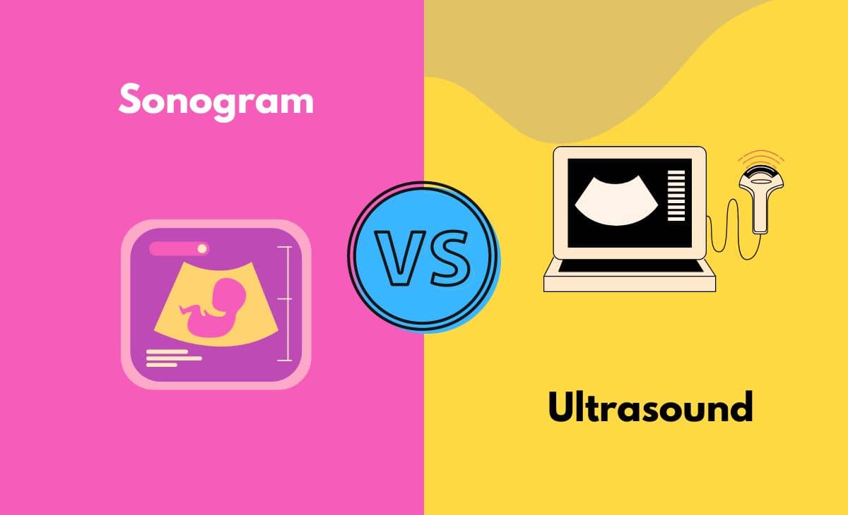
The main difference between an ultrasound and a sonogram is that an ultrasound refers to a procedure, while a sonogram is the image that is created by that process. Sonogram, in layman's terms, does not refer to the procedure itself; rather, it is the exact consequence of another technique called ultrasound, which is sometimes confused with the same process. It should be obvious that an ultrasound is a procedure; it is a form of imaging test, and the output that it produces is called a sonogram.
Sonogram, which is also known as sound writing, is the output that is produced by ultrasound, which is also known as diagnostic sonography. In other words, a sonogram is a sound writing. This technology is very helpful in evaluating whether or not there is an infection or disease present in the interior sections of the body, which primarily includes the organs and tissues found within the body.
Sonography is favored over other imaging procedures because of the lower risk of complications it presents. This technique does not involve the use of any radiation. The patient is subjected to considerable doses of radiation whenever they go through the process of getting a CT scan done. In the case of MRI, pictures are obtained with the assistance of magnets that have an exceptionally high field strength. Patients who have metal implanted in their bodies cannot undergo MRI testing.
The sonography examination is performed on the outermost layer of the patient's skin, and there are no known risks or side effects related with the procedure. It is generally agreed that the ultrasound waves are completely safe. There is a possibility that the integrity of tissues will be compromised if they are subjected to ultrasonic waves for an extended period of time. However, the computer modifies the strength of the sound waves, and the sonographer employs several methods to cut down on the amount of time spent in exposure. As a result, sonography is considered to be a comparatively safe imaging procedure in comparison to others.
Difference Between Sonogram and Ultrasound in Tabular Form
| Parameter of Comparison | Sonogram | Ultrasound |
| Definition | The final product of one operation is referred to as a sonogram, which is a graphical representation of the data obtained from that process. | The process of producing images of the interior of the body through the use of soundwaves is referred to as ultrasound. |
| Result vs Process | The final product of an ultrasound is called a sonogram. | Sonograms can be created by a process called ultrasound, which describes the method. |
| Relation to Sonography | Sonogram is the technical term for the visual image that is produced by sonography. | Sonography is carried out with the use of an ultrasound device. |
| Motive | In cases of pregnancy, a sonogram can be used to generate images of the developing baby and assist in the evaluation of organs for infections, injury, or disease. | Sonograms, which provide diagnostic physicians with insights into the inner workings of a human body, can be obtained with the assistance of ultrasound technology. |
| Alternatively called | It is also sometimes referred to as sound writing,' which is another name for it. | The term "Diagnostic Sonography" is another name for this procedure. |
What is a Sonogram?
Sonography is the technique of producing a visual image through the use of sound waves, and the resulting image is referred to as a sonogram. When the sound waves come into contact with a surface, they reflect off of it and then bounce back, which is what causes the visuals to be generated.
To provide further context, the sound waves reflect back an increasing amount the more compact and difficult the surface is. For example, sound waves can easily travel through liquids, and because of this, they will produce an image that is completely black when they come into touch with liquids such as urine, water, or other liquids.
In addition, when they strike a tissue, the strength of the sound waves causes them to produce an image that is either dingy or bright, depending directly on the color of the tissue. In addition, when they come into contact with extremely dense tissues or tougher substances, such as the bones or the kidneys, it is evident that they will show up as a brighter white area in the picture.
Sonography is a diagnostic medical test that creates an image of the inside of the body by using high-frequency sound waves, which are sometimes referred to as ultrasound waves, and bouncing them off of various structures. This diagnostic procedure is also known as an ultrasound and a sonogram, which is only fitting.
Sonography works by sending ultrasonic waves into the body and listening for an echo using a device called a transducer that is placed on the surface of the patient's skin. An image is produced as a result of the ultrasonic waves being translated by a computer. Structures in the image are visible to a qualified technician, who can also measure and identify them. The photos are then read by a medical professional to assist in making a diagnosis of the issue or condition at hand.
Sonograms take real-time pictures of what's happening on the interior of a patient's body. It works much like a camera and can take photographs of different body parts or processes while they are happening in real time.
Sonography can be helpful in the diagnostic process for a variety of medical disorders since it can evaluate the size, shape, and density of tissues. Ultrasound imaging has traditionally been considered an excellent method for examining the abdominal region without the necessity of making an incision.
Recently, sonograms have proven to be of significant value in the field of medicine. They have been an extremely important factor in the development of a basis for evaluating the internal organs of the body, as well as in the diagnosis and ultimate treatment of infections and disorders that originate in the body's internal organs or its soft tissues.
What is Ultrasound?
Sonograms can be produced by a technique known as ultrasound, which employs the utilization of sound waves. In the realm of medical diagnostics, you can think of it as an image examination. They do not use any kind of radiation that could be hazardous; rather, they make use of high-frequency sound waves, which are neither uncomfortable nor hazardous to the user.
When the sound waves are reflected back, they produce electrical signals. These electrical signals are then translated by a computer to provide images of the various tissues and organs found within the body. In order to obtain a clearer picture during an ultrasound, a transducer may be placed on the surface of the skin or it may be implanted into one of the body's natural openings. Both of these methods are utilized in the diagnostic process.
Sonography and ultrasonography are both terms that refer to ultrasound, which is a noninvasive imaging test. A sonogram is the picture that is produced by an ultrasound. Creating images or video in real time of internal organs or other soft tissues, such as blood vessels, can be accomplished with the use of ultrasound by employing high-frequency sound waves.
The use of ultrasound technology gives medical professionals the ability to "see" the structure of internal soft tissues without having to make any wounds (cuts). Additionally, in contrast to X-rays, ultrasound does not make use of any radiation.
Even though most people only think of ultrasounds being used during pregnancies, doctors and other medical professionals utilize them for a wide variety of diagnostic purposes and to examine many different organs and sections of the human body's interior.
The specialized transducer could be inserted into the body in one of three primary ways: through an ultrasound, which entails placing the transducer; through a transrectal ultrasound, which entails placing the transducer; or through a transoesophageal echocardiogram, which entails placing the transducer in the oesophagus. All three of these methods involve placing the transducer.
In most cases, ultrasound is utilized to assist in the diagnosis of diseases that are related to the internal organs and soft tissues of the body. Additionally, ultrasound is widely renowned for its ability to both confirm and monitor pregnancy.
Main Differences Between Sonogram and Ultrasound in Points
- The procedure of ultrasound results in the creation of a graphical representation known as a sonogram.
- An ultrasound makes use of soundwaves to generate electrical signals, which are then transformed by a computer into visual pictures known as sonograms. These sonograms can be used to diagnose a variety of medical conditions.
- Sonograms, which are produced as a result of ultrasound, can be used to assist in the diagnosis of illnesses and infections that need to be treated. This is made possible by the fact that ultrasound makes it possible to view images of the interior organs of the body.
- The handheld transducer, computer, monitor, and printer that make up an ultrasound system are used to perform the procedure, but the computer alone is responsible for creating sonograms from the electrical signals that are received.
- Sonograms are visual photographs that can also be printed in a physical manner, but ultrasound is a delicate procedure that uses a variety of instruments such as a transducer and other similar devices.
- The terms "ultrasound" and "sonography" are frequently used interchangeably; nevertheless, ultrasound refers specifically to the process of employing sound waves to create images from inside the body. The image that is produced by an ultrasound examination is called a sonogram.
Conclusion
Sonogram and ultrasound, two terms that are frequently substituted for one another, are actually components of the same procedure. However, each of these phrases can be understood in two completely distinct ways on their own. It would be erroneous to consider them to be ordinary medical terminology because doing so would remove the requirement to understand them.
When you proudly present the printout to all of your friends and family members, there are some helpful hints that can be used to figure out what it is that you are actually looking at. What should we most take away from this? The parts that are black represent the amniotic fluid, whereas the ones that are gray represent the soft tissue. Even if ultrasound technology is improving every day — for instance, the doctor might offer a 3D version — it may still be difficult to make out the subtleties of the image. Therefore, in order to orient oneself, you should attempt looking for the limbs or the head.
As a result of the fact that these procedures are utilized extensively in the modern medical system and are performed on a regular basis, it is essential for the average person to be aware of and comprehend them in the event that they are subjected to one of these tests.
It is imperative that we comprehend the fact that these technologies have been developed in order to assist both ourselves and the medical system in locating treatments for ailments that are more complex and technical in nature.
References
- https://medlineplus.gov/lab-tests/sonogram/
- https://medlineplus.gov/ultrasound.html

