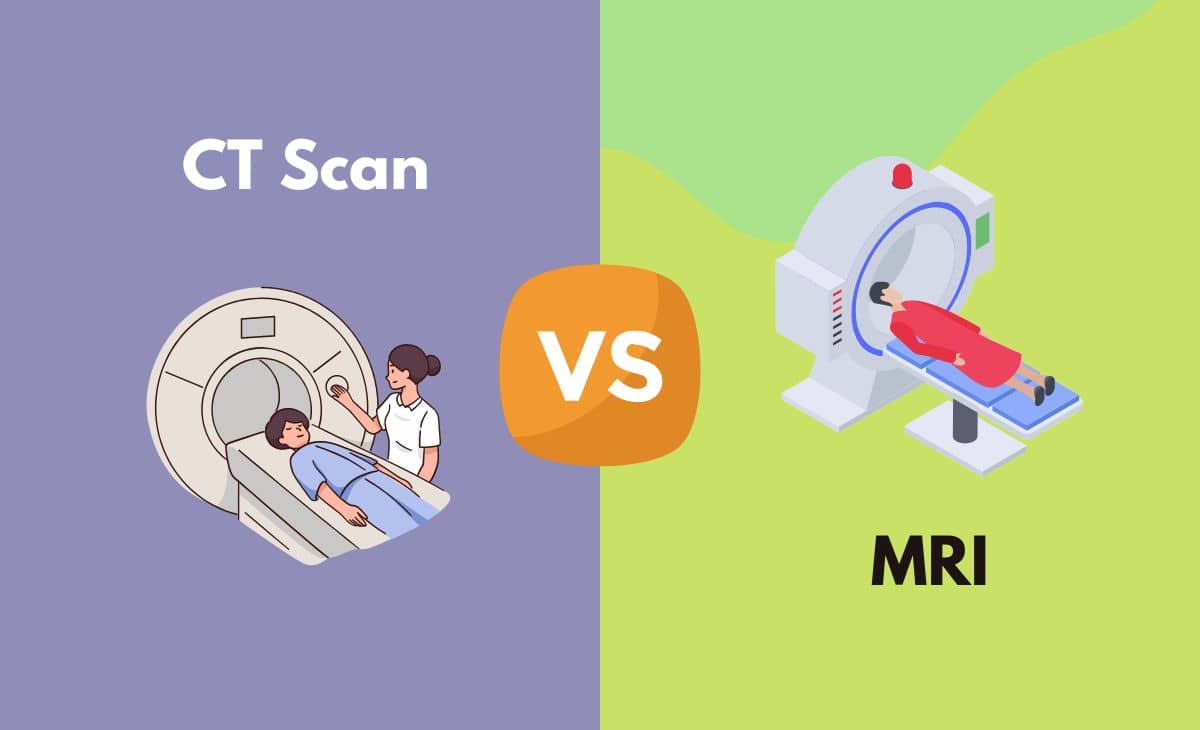
The terms CT scan (CAT scan) and MRI is known to everyone living in the modern world. It's interesting to note that these devices may mean different things to different people. A layperson views them as devices used in hospitals when doctors cannot make an immediate diagnosis and want to do a thorough search. In the case of hospital employees (doctors, nurses etc.), the CT scan and MRI are devices used to identify the exact cause of the medical problem. On the other hand, for an airport security worker, a CT scan is used to scan the luggage of people travelling by aeroplanes.
Computed (axial) tomography and magnetic resonance imaging devices are used as medical imaging techniques. In the hospital setting, they are used to develop detailed pictures of internal body parts (bones, organs, joints, etc.)
While most people may know of the devices, they may not be aware of the differences between them or even why one of the devices gets picked over the other.
CT Scan vs MRI
Both are apparatuses used for producing images of the internal structures of the human body.
A CT scan uses X-rays to produce images of a human body or any object that can pass through the machine. Mostly it is used to scan for bone fractures, detect internal bleeding, scan for tumours, or to detect cancer. It is an expeditious procedure and is completed within five minutes.
An MRI machine, on the other hand, can use radio waves and extremely powerful magnets to produce images of the inside of the human body. It is generally used to scan for soft tissue damage or spinal cord injuries. It is a lengthy procedure and may take up to 20 or 120 minutes depending on the need of the scan. Compared to a CT scan, it is also a costly procedure.
Difference Between CT scan and MRI in Tabular Form
| Parameters of Comparison | CT Scan | MRI |
| Full form | Computed (Axial) Tomography | Magnetic Resonance Imaging |
| Radiation | Present | None |
| Time taken | 5 minutes | Depending on the need and area of search either takes 15 minutes or 2 hours |
| Application | Emergency room patients, bone injuries, cancer detection, lung and chest imaging | Brain tumours, soft tissue evaluation, spinal cord injury |
| Cost | Less expensive than MRI | Expensive |
| The method used for scanning | X-rays | Radio waves |
| Noise factor | Quiet | Noisy |
| Main disadvantage | Exposure to radiation can cause cancer | Causes extreme distress for person with claustrophobia |
What is a CT scan?
The computer (Axial) Tomography was invented by Sir Godfrey Hounsfield in 1973, an invention for which he also won a Nobel Prize. It is a device which is used to scan substances such as the human body. According to Merriam-Webster’s dictionary, computed tomography means, radiography in which a 3-D picture of an object is fabricated by a computer from a string of plane cross-sectional images constructed along an axis. In simple terms, it is a big X-ray machine in which a person can lie down.
History
Gabriel Frank (1940) in one of his patents put forth a rudimentary idea for tomography. His patent contained diagrams for a piece of equipment to form sinograms and optical backprojection approaches to reconstruct images. The images produced by this approach were blurred.
It wasn’t until twenty-one years had passed, that further improvements were seen. William H. Oldendorf, a neurologist stemming from Los Angeles executed a series of experiments to ascertain whether internal structures enclosed by dense structures could be found out by the use of transmission measurements. The principles he used for this work are similar to the ones later used in computer tomography.
Two years later, David E. Kuhl along with Roy Q. Edwards developed transverse tomography by utilizing radioisotopes. The further development of this technique led to modern emission computed tomography.
In the year 1963, Allan M. Cormack presented the results from investigations into the first CT scanner ever developed.
In 1967, Godfrey N. Hounsfield started the work on the first CT scanner, at the central Research Laboratories. Godfrey was exploring pattern recognition techniques, when he extrapolated that if the X-ray images of an object were taken from variable directions, it would help in the reconstruction of its internal structure.
In September 1971, the first Computer tomography device was in Atkinson-Morley Hospital, London. In the October of 1971, the device scanned its first patient and the anatomy of the patient was distinctly visible.
In 1979, Cormack and Hounsfield received the Nobel Prize in Physiology and Medicine for their in Computed tomography.
Procedure of CT Scan
A CT scan utilizes X-rays to create intricate images of bones, organs etc. The patient will be made to lie down on a table. The table will then move through a scanning circle, which is the CT scanner. The CT scan will then take cross-sectional images of the inside of the patient's body. The data collected from this procedure is constructed to form 3-D images. These images will reveal if there are any abnormalities in the bone or soft tissue structures or if the person has any tumours etc.
Uses of a CT scan
In addition to human bodies, a CT scanner can be utilized to scan animals, mummies, trees, industrial parts, scan baggage in an airport, etc. In short, it can be used to scan essentially anything which can be placed inside a CT frame.
In a medical setting, it is used for
- Scanning bone fractures
- Detecting internal bleeding
- Scanning for tumours
- Cancer monitoring
Workings of the CT scan
In general, the CT scan uses X-rays to create pictures of the human body or any objects that will go into the scanner. Inside the CT scanner, the X-ray tube will revolve around the person/ object lying inside. In the opposite direction of this X-ray tube, is the X-ray detector. The X-ray detector will receive the emission that makes its way through the person. This emission is then sampled via approximately 764 channels. The signals acquired by each channel are then digitized to a 16-bit value.
Afterwards, it is transferred to the reconstruction processor. The measurements taken by the CT scanner are around 1000 times per second. The scan rotations usually take about 1 to 2 seconds. Every channel piece of scanned information is then compared to a calibration scan material of air, polyethene and water. These materials were previously acquired in a comparable location. These comparisons will allow the picture element to possess a known value for a specific substance in the human body. The more picture samples it takes, the better the final image will be.
Advantages of CT Scan
- Images of the entire body can be developed.
- Less time consuming
- Exceptionally useful for diagnosing cancer and its stage. It is also used to check if cancer has returned.
- Used to monitor if treatments are working properly.
- Good for producing pictures of bone structures.
- Cheaper than an MRI
Disadvantages of CT Scan
- A CT scan uses radiation, and repeated exposure to this can cause cancer in patients.
- The CT scan makes use of ionizing radiation. This could harm DNA.
- It is harmful to pregnant women and their unborn children.
- The CT scan sometimes uses a specific dye known as contrast material. This can cause negative reactions in some patients such as headache, nausea, itching, vomiting, skin rash, fever and musculoskeletal pain.
What is MRI?
MRI is an acronym for Magnetic Resonance Imagery. It is a type of image-scanning device that uses radio waves and powerful magnets to fabricate pictures.
Procedure of MRI
As with a CT scan, an MRI machine also requires that the person lies inside it. The table will move inside a doughnut-shaped device, which is the MRI scanner. The MRI scanner then creates a constant magnetic field and makes use of radio waves to deflect water molecules and fat cells inside the human body. The radio waves inside the machine are relayed to a receiver in the MRI apparatus. This is then translated to images of the human body for diagnostic purposes. The pictures taken with an MRI machine can reveal the difference between normal tissue and diseased tissue.
An MRI machine is typically loud, as a patient, you will be offered earplugs or headphones before going in the machine, to help make the noise tolerable.
Uses of an MRI Machine
An MRI machine makes use of radio waves and powerful magnets to develop images that can display the inside of a body. Majorly, they are used to help diagnose problems with,
- Joints
- Ankles
- Brain
- Breasts
- Wrists
- Heart
- Blood vessels etc.
Workings of an MRI Machine
The MRI machine uses extremely powerful magnets and pulsing radio waves. The detection coils present inside the MRI scanner wade through the energy created by the water molecules as they reorganize themselves after each RF alignment pulse.
Afterwards, the data collected from the scanner is recreated into a two-dimensional image through any axis of the body. The bones of living organisms are practically empty of water. Hence, they do not give rise to any images. This leads to the presence of a black area in the pictures. An MRI machine is best applicable to capture images of soft tissue.
Advantages of an MRI Machine
- Provides more intricate images of soft tissues
- Can change the contrast of the pictures produced
- Can change the imaging plane without affecting the patients’ position.
- Contrast agents utilized in MRI are safer than the ones used in X-rays.
- MRI is the superior equipment for detecting tumours and identifying them.
- Certain cancers like prostate cancer, liver cancer and uterine cancer cannot readily be detected using a CT scan. Hence in these cases, an MRI is better suited.
Disadvantages of an MRI Machine
- An MRI machine requires that the patient lay still in the machine for 20 – 40 minutes. This can cause distress to people with claustrophobia.
- The procedure is noisy. This can cause hearing issues.
- Risk of possible negative reaction to metals, as the machine uses magnets.
- During long-duration MRI, it can cause an increase in the patient's body temperature.
In addition, it is wise to consult a doctor before taking an MRI if you have any of the following implants:
- Artificial joints
- A pacemaker
- Eye implants
- An IUD
Main Differences Between CT scan and MRI in Points
- CT scan is an acronym for computer (axial) tomography. MRI is an acronym for magnetic resonance imagery.
- A CT scanner uses radiation. An MRI does not use radiation.
- The MRI machine is very noisy and the patients need to be provided with earplugs for the noise to be tolerable. A CT scan in comparison is very quiet.
- A CT scan is very quick; it usually takes only up to 5 minutes. An MRI machine on the other hand can either take up to 15 minutes or 2 hours.
- A CT scanner uses X-rays for capturing images. In contrast, an MRI machine uses radio waves and powerful magnets.
- A CT scan is usually used for emergency room patients. An MRI is used to find more complicated problems.
- Images from a CT scan are used to find bone injuries, cancer, internal bleeding or tumours. An MRI machine is used to scan for problems in joints, the brain, ankles, breasts, the heart, etc.
- Comparatively, a CT scan is less expensive than an MRI scan.
How a Choice Between the CT Scan and MRI is made?
Since both the devices are similar doctors will usually choose one machine over the depending upon a variety of factors,
- Pregnancy
- The medical purpose of the scan
- Level of intricacy required from the image
- Claustrophobic patient
- An MRI is chosen when a precise image of organs, soft tissues or ligaments is needed. Some example cases are torn ligaments, herniated disks, soft tissue damage, etc.
- A CT scan may be chosen over an MRI for a generalized picture of internal organs, or in cases with fractures or head traumas.
Conclusion
In conclusion, a CT scan and an MRI are both procedures used to get an image of the inside of the body. While they are similar in some ways, they both produce the information necessary for a diagnosis and they are also relatively low-risk procedures. A CT scan uses X-rays for imagery. It is used to scan a general or large area. MRI uses radio waves and magnets. It is used to develop a superior image of a tissue sample. The choice between the two will be made by a doctor.
References
- Hsieh, J. (2009). Computed tomography: Principles, design, artifacts, and recent advances (2nd ed). Wiley Interscience ; SPIE Press.
- https://www.merriam-webster.com/dictionary/computed%20tomography
- https://www.mskcc.org/news/ct-vs-mri-what-s-difference-and-how-do-doctors-choose-which-imaging-method-use
- https://www.medicalnewstoday.com/articles/326839

